The Retina

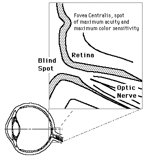
The retina is a light-sensitive layer at the back of the eye that covers about 65 percent of its interior surface. Photosensitive cells called rods and cones in the retina convert incident light energy into signals that are carried to the brain by the optic nerve. In the middle of the retina is a small dimple called the fovea or fovea centralis. It is the center of the eye's sharpest vision and the location of most color perception.
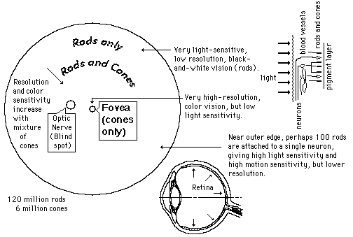
"A thin layer (about 0.5 to 0.1mm thick) of light receptor cells covers the inner surface of the choroid. The focused beam of light is absorbed via electrochemical reaction in this pinkish multilayered structure. The human eye contains two kinds of photoreceptor cells; rods and cones. Roughly 125 million of them are intermingled nonuniformly over the retina. The ensemble of rods(each about 0.002 mm in diameter in some respects has the characteristics of a high-speed, black and white film(such as Tri-X). It is exceedingly sensitive, performing in light too dim for the cones to respond to, yet it is unable to distinquish color, and the images it relays are not well defined."(Hecht)

"In contrast, the ensemble of 6 or 7 million cones (each about 0.006 mm in diameter) can be imagined as a separate, but overlapping, low-speed color film. It performs in bright light, giving detailed colored views, but is fairly insensitive at low light levels."(Hecht)"

Index

Vision concepts

Image formation concepts

Reference
Hecht, 2nd Ed.
Sec. 5.7
 
HyperPhysics***** Light and Vision R Nave
Go Back


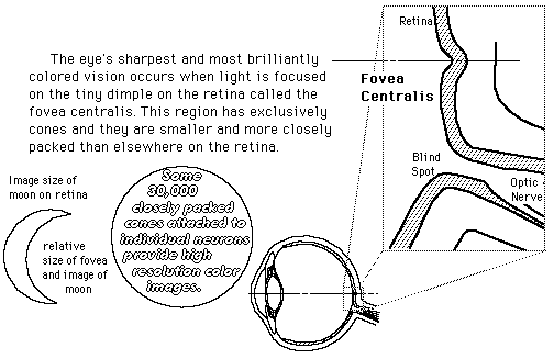
The Fovea Centralis

Though the eye receives data from a field of about 200 degrees, the acuity over most of that range is poor. To form high resolution images, the light must fall on the fovea, and that limits the acute vision angle to about 15 degrees. In low light, this fovea constitutes a second blind spot since it is exclusively cones which have low light sensitivity. At night, to get most acute vision one must shift the vision slightly to one side, say 4 to 12 degrees so that the light falls on some rods.

A "dimple on the retina" provides our highest resolution vision.

"Just about at the center of the retina is a small depression from 2.5 to 3 mm in diameter known as the yellow spot, or macula. There is a tiny rod-free region about 0.3mm in diameter at its center, the fovea centralis. (In comparison the image of the full Moon on the retina is about 0.2 mm in diameter.) Here the cones are thinner (with diameters of 0.0030mm to 0.0015mm) and more densely packed than anywhere else in the retina. Since the fovea provides the sharpest and most detailed information, the eyeball is continuously moving, so that light from the object of primary interest falls on this region. ...the rods are multiply connected to nerve fibers, and a single such fiber can be activated by any one of about a hundred rods. By contrast, cones in the fovea are individually connected to nerve fibers. The actual perception of a scene is constructed by the eye-brain system in a continuous analysis of the time-varying retinal image."(Hecht)

Index

Vision concepts

References
Hecht, 2nd Ed.
Sec. 5.7
Williamson & Cummins
 
HyperPhysics***** Light and Vision R Nave
Go Back


Optic Nerve

The optic nerve is the cable of nerve fibers with carries the electrical signals from the retina to the brain for processing. The point of departure of that optic nerve through the retina does not have any rods or cones, and thus produces a "blind spot".

Index

Vision concepts

References
Hecht, 2nd Ed.
Sec. 5.7
Williamson & Cummins
 
HyperPhysics***** Light and Vision R Nave
Go Back