The Heart's Electrical Sequence
The synchronized electrical sequence of the heart is initiated by the SA node, the heart's natural pacemaker. The firing of the SA node sends out an electrical impulse via its neurons to the right atrium, left atrium, and AV node simultaneously. Since the right atrium is closer to the SA node, it depolarizes first, resulting in pumping action by the right atrium before the left atrium. At the AV node, the impulse is delayed to allow for the ventricles to fill up with blood. After the delay, the AV node sends the impulse to the Bundle of His and the Purkinje fibers. This triggers the contraction of the ventricles to send blood either to the lungs or out to the body.
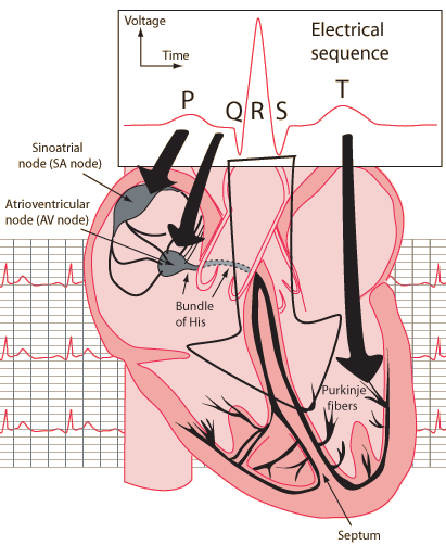
| P Wave | Firing of the SA node and depolarization of the atria. |
| PR Interval | Delay of the electrical impulse at the AV node and the depolarization of the atrium. |
| QRS Complex | Ventricular depolarization
|
| ST Segment | The beginning of ventricular repolarization. Should be isoelectric (flat at baseline). |
| T Wave | Ventricular repolarization. |
| Electrocardiograms |
Bioelectricty
References
Chernecky
Grauer
| HyperPhysics***** Biology | R Nave |
